About OCT
Optical Coherence Tomography (OCT) technology is a high speed, high definition (HD), and noninvasive imaging technology used to obtain high resolution cross-sectional images of the retina. With OCT imaging, the individual layers of the retina can be differentiated and retinal thickness can be measured. This is very useful for the early detection and diagnosis of retinal diseases and conditions such as macular degeneration, glaucoma, and diabetic retinal disease.

About Optomap
The Optos Optomap® provides an ultra-widefield, high resolution image of the retina. This provides a much broader and more detailed view of the retina than is possible with conventional imaging methods. Our doctors strongly believe that the Optomap® Retinal Exam is an essential part of your comprehensive eye exam and prescribes it for all patients once per year. One of the many benefits of this technology is that it can serve as an alternative to routine dilation of your eyes; however, dilation may still be required to more closely evaluate the retina if abnormalities are detected on screening images.
Optomap is an innovative new technology that gives eye doctors the ability to perform ultra-wide retinal imaging that is far superior to what can currently be achieved using conventional retinal imaging options. In contrast to conventional retinal imaging, Optomap captures at least 50% more of the retina in a single capture, and with Optomap’s multi-capture function, up to 97% of the retina can be viewed. This gives eye care professionals greater opportunity to monitor the health and condition of patient vision.
Why Is Optomap Important?
Optomap is another great preventative eyecare technology tool. By allowing your eye doctor to have a comprehensive view of your retina, they will be able to detect any developing eye diseases early on, before they have a detrimental impact on your vision and day to day life. Not only can Optomap detect eye conditions such as retinal holes, retinal detachment, macular degeneration and diabetic retinopathy, but it can also be used to identify some general health conditions such as cardiovascular disease, stroke and cancer.
What To Expect From Optomap Scanning
Optomap is a fast, painless and non-invasive procedure that is suitable for patients of all ages, even children and pregnant women. Many patients require their eyes to be dilated ahead of the scan and will be given eyedrops which will widen their pupils and make it easier for the camera to see the structures inside the eye. Pupil dilation is painless, but patients may feel more sensitive to light both during their Optomap scan and afterwards for up to 24 hours. You may also have slightly blurred vision for a few hours. Once your eyes are dilated, you’ll be sat down and asked to look into a small device that will take the pictures of your retina. A short flash of light will let you know that the image has been taken, and the entire imaging is over in just a few seconds. The results will be sent digitally to your eye doctor who will then evaluate them. The results will also be stored on your personal optical record for future information.
If you would like more information about what is involved in Optomap, or to schedule an appointment for this effective screening technology, please contact our eyecare team.

About icare ONE
The iCare tonometer is a handheld, minimally invasive device that measures the eye’s intraocular pressure without requiring any eye drops. Measuring the eye’s intraocular pressure is an important part of any eye examination because it allows us to detect the presence of ocular diseases such as glaucoma.

About Diopsys
The Diopsys® RETINA PLUS™ Vision Testing System administers painless, non-invasive Light Induced Visual-response (LIV)™ tests to provide your doctor with comprehensive objective information about how well your visual system is functioning.
Like an electrocardiogram (EKG) which tests heart function, LIV tests work by evaluating how the cells within your vision system are functioning. Many eye diseases disrupt normal cell function as they become unhealthy. By catching this dysfunction before the cells die, your doctor may be able to prescribe treatment to make the cells healthy again. Additionally, your doctor can use the tests to help determine if your treatment is working.
This technology has traditionally been used to diagnose rare eye diseases, but is now able to help manage and diagnose more common eye diseases like: diabetic retinopathy, glaucoma, optic neuritis, and more.

About iLUX
The iLUX® Device is used to heat and compress glands in the eyelids of adult patients with a specific type of dry eye, called Meibomian Gland Dysfunction (MGD), also known as evaporative dry eye. Potential side effects may include eyelid/eye pain requiring stopping the treatment procedure, eyelid/eye irritation or inflammation, temporary reddening of the skin, and other eye symptoms (burning, stinging, tearing, itching, discharge, redness, feeling like there is something in the eye, changes in your vision, sensitivity to light). Ask your eye care professional for a complete list of safety information for the iLUX® Device."
What is Intensed Pulse Light Treatment?
Intense Pulsed Light or IPL treatment is a skin treatment that improves dry eyes. IPL treatment has been used by Dermatologists for over twenty years. For example, people who had IPL treatment for Rosacea found their dry eyes were significantly improved. IPL can directly deal with one of the leading causes of dry eyes known as Meibomian Gland Dysfunction. MGD is the leading cause of over 86% of dry eye cases along with other factors. This dysfunction occurs when the meibomian glands don’t produce enough oil for the tears which keeps the eye moist.
How Can IPL Help With Dry Eyes?
IPL treatment involves flashes of light to the skin around the eyelids and face. Light gets absorbed by the mitochondria of the Meibomian Glands. This helps to switch on the Glands. They become 'younger' and more active. The Glands produce better Meibomian Oil and improve dry eyes. IPL also helps target Ocular Rosacea. Flashes gets absorbed in the small, leaky capillaries in Ocular Rosacea. It helps to ‘seal off’ these leaky vessels. The result is less inflammation and redness. IPL also kills bacteria and Demodex on the surface of the eyelids. In short, IPL helps switch on your eyes and break the vicious cycle of dry eyes.

OCULUS Keratograph® 5M
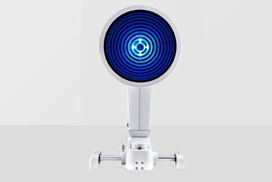
The OCULUS Keratograph® 5M is an advanced corneal topographer and color imaging system optimized for capturing images of the external eye and its surrounding structures. Unique features include the ability to evaluate dry eye and capture high resolution photos and videos of the anterior segment (front portion) of the eye. It is also very helpful in the fitting of contact lenses and the diagnosis of corneal disorders such as keratoconus.

Humphrey Automated Visual Field
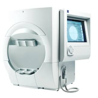
The Humphrey Field Analyzer 3 is a subjective / patient-interactive test that is used to determine an individual's visual sensitivity. Results from this test are used to determine if there is an abnormality within the eye or visual pathway. Certain diseases that can be detected with this device include glaucoma, stroke, traumatic brain injury, toxic injury to the retina or optic nerve, and stroke (cerebrovascular accident).

Optilight
OptiLight by Lumenis is a safe, gentle, and effective treatment done to manage dry eye disease.
This non-invasive procedure is the first and only FDA-approved intense pulsed light (IPL) treatment for dry eye management.
OptiLight uses pulses of light precisely administered in the area below the eyes to reduce dry eye symptoms. This 10-15 minute procedure can relieve dry eye symptoms by:
- Increasing tear break-up time
- Reducing the amount of demodex mites and bacteria around your eyes
- Decreasing inflammation inflammation
- Improving meibomian gland functionality


